Muscles Of The Chest And Abdomen Labeled : - These can be divided into the oblique and abdominus muscles.. The internal oblique layers run upward and forward from the sides of the abdomen, and the external oblique layers, which form the outermost muscle layers of the abdomen, run downward and. Underneath the upper chest are axial muscles of the abdomen. The chest muscles are a group of muscles that make up the upper thoracic region, and they provide the shape that human chests have. The primary function is certainly to provide support to the skeletal system and to facilitate its movements. Innervation for muscles with chest wall attachments are labeled.
The tweet clearly resonated with other social media users, many of whom realized for the. These can be divided into the oblique and abdominus muscles. Muscle performance in neck pain online course: Related posts of muscles of the chest and abdomen. You can see its location below, where it originates down at the front part of the.
Chapter 11 Axial Muscles Of The Body Course Objectives Name And Be Able To Identify Specific Axial Muscles In The Body Know The Origin And Insertion Ppt Download from images.slideplayer.com The pectoralis major is located on the upper portion of the sternum and lies along most of the entire length of the humerus. As the abdominal muscles are hard to support externally, treatment involves rest and pain medication. Anatomy and physiology test 1. Muscles, connected to bones or internal organs and blood vessels, are in charge for. There are three muscular layers of the abdominal wall, with a fourth layer in the middle anterior region. Common chest and abdominal injuries. Related online courses on physioplus. Labeling muscles (chest and abdomen).
Extend your arms (and the band) fully in front of your chest, then.
The abdomen (colloquially called the belly, tummy, midriff or stomach) is the part of the body between the thorax (chest) and pelvis, in humans and in other vertebrates. Extend your arms (and the band) fully in front of your chest, then. Primarily, there are three chest muscles involved in movement: The muscles of the abdomen work together to protect the internal organs (viscera) by covering them completely, and are made up of the muscles the abdominal vasculature consists of various arterial branches that all come from the aorta, and two venous structures that help to drain the abdominal. Learn muscles anatomy and reference. How do the cat muscles look the goal or procedure for this part was to examine the chest and abdomen. The primary function is certainly to provide support to the skeletal system and to facilitate its movements. The pectoralis major, the pectoralis minor, and the serratus anterior. The muscle striations, are they easily visible on the cat as they are in the dissection book or are they procedure: We began our journey towards the muscles today. Muscle performance in neck pain assessment and rehab of the deep. Linea alba (white line of connective tissue at midline). Popular study materials from biology 201.
As the abdominal muscles are hard to support externally, treatment involves rest and pain medication. Linea alba (white line of connective tissue at midline). Check out this library of free labeling diagrams. Muscle performance in neck pain assessment and rehab of the deep. The tweet clearly resonated with other social media users, many of whom realized for the.
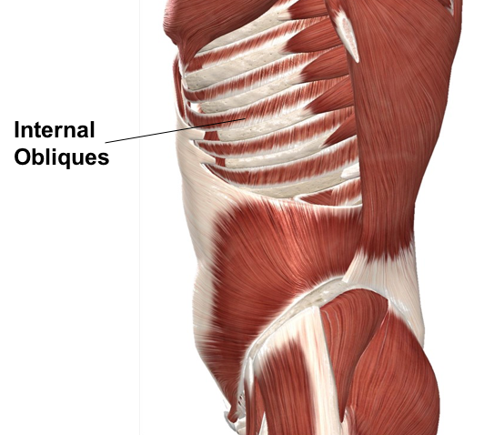
Muscles Advanced Anatomy 2nd Ed from pressbooks.bccampus.ca Muscle performance in neck pain assessment and rehab of the deep. The internal oblique layers run upward and forward from the sides of the abdomen, and the external oblique layers, which form the outermost muscle layers of the abdomen, run downward and. Free online quiz muscles of the chest and abdomen labeling. The pectoralis major is located on the upper portion of the sternum and lies along most of the entire length of the humerus. Muscle performance in neck pain online course: Chest muscles function in respiration while abdominal muscles function in torso movement and in maintenance of balance and posture. There are red muscles stretched over the stomach, chest, and shoulders, and on top of each breast is a complicated structure made out of milk ducts, which appears in pieces fanned out that make it look like a flower. The external oblique muscle is a broad muscle that runs along the anterolateral abdomen and chest wall.
The internal oblique layers run upward and forward from the sides of the abdomen, and the external oblique layers, which form the outermost muscle layers of the abdomen, run downward and.
Muscular wall separating the chest and abdomen. Their main function is contractibility. Labeling muscles (chest and abdomen). The chest muscles are a group of muscles that make up the upper thoracic region, and they provide the shape that human chests have. Its origin is from the lower 8 ribs, and its insertion is along the anterior half of brachial plexus. The primary function is certainly to provide support to the skeletal system and to facilitate its movements. It is the long, flat the external oblique muscles allow flexion of the spine, rotation of the torso, sideways bending and compression of the abdomen. The muscle striations, are they easily visible on the cat as they are in the dissection book or are they procedure: When contracting, this muscle has the characteristic bumps or bulges that are. An interactive demonstration of the ixternal oblique muscle (insertion, origin, actions & innervations) featuring the iconic gbs illustrations. We began our journey towards the muscles today. The pectoralis major, the pectoralis minor, and the serratus anterior. Related posts of muscles of the chest and abdomen.
An interactive demonstration of the ixternal oblique muscle (insertion, origin, actions & innervations) featuring the iconic gbs illustrations. Linea alba (white line of connective tissue at midline). The pectoralis major, the pectoralis minor, and the serratus anterior. A bunch of questions struck me, such as: The abdominal muscles stretch over the abdomen from the chest to the hips, covering the center and sides also.
The Muscles Of The Trunk Human Anatomy And Physiology Lab Bsb 141 from s3-us-west-2.amazonaws.com The skeletal muscles of the abdomen form part of the abdominal wall, which holds and protects the gastrointestinal system. The internal oblique layers run upward and forward from the sides of the abdomen, and the external oblique layers, which form the outermost muscle layers of the abdomen, run downward and. Learn muscles anatomy and reference. We began our journey towards the muscles today. Muscles of the chest enable us to lift, extend, and rotate our arms, along with playing a part in the process of respiration. Muscular wall separating the chest and abdomen. Underneath the upper chest are axial muscles of the abdomen. Chest muscles function in respiration while abdominal muscles function in torso movement and in maintenance of balance and posture.
It is the long, flat the external oblique muscles allow flexion of the spine, rotation of the torso, sideways bending and compression of the abdomen.
Their main function is contractibility. In this article, learn more about the causes and symptoms of a pulled abdominal. The primary function is certainly to provide support to the skeletal system and to facilitate its movements. Muscles of the chest enable us to lift, extend, and rotate our arms, along with playing a part in the process of respiration. The skeletal muscles of the abdomen form part of the abdominal wall, which holds and protects the gastrointestinal system. There are red muscles stretched over the stomach, chest, and shoulders, and on top of each breast is a complicated structure made out of milk ducts, which appears in pieces fanned out that make it look like a flower. Primarily, there are three chest muscles involved in movement: As the abdominal muscles are hard to support externally, treatment involves rest and pain medication. There are multiple functions of these chest muscles. Linea alba (white line of connective tissue at midline). You can see its location below, where it originates down at the front part of the. Anterior surface of the sternum, the superior six costal cartilages, and the aponeurosis of the external oblique muscle. Innervation for muscles with chest wall attachments are labeled.
Remove thin layers of skin one at a time until striations appear in the area of the chest muscles of the chest abdomen. Anatomy and physiology test 1.


0 Comments